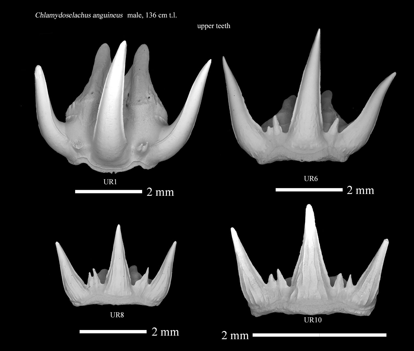
| A study on the striae of the frilled shark Chlamydoselachus anguineus Garman, 1884 was previously reported on this site (Nakagawa, 2012*1). In this study, we examined the striae in more detail. The teeth examined were selected from a dentition isolated from a 136 cm long male shark caught in Tokyo Bay. As a result, each of the examined teeth showed distinct differences in the morphology of the striae due to differences in their original position in the dentition. Based on these results, we estimated the growth process of striae in the teeth of the frilled shark. |
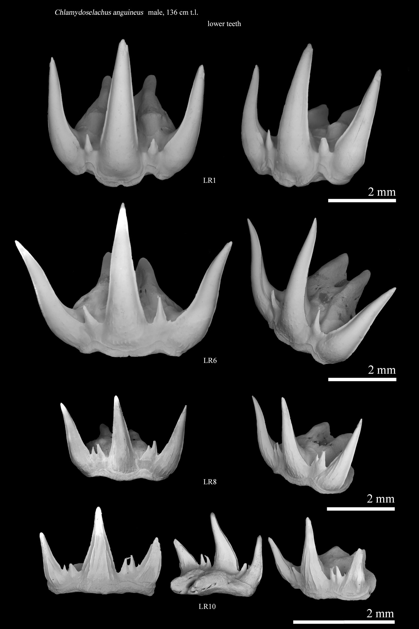
| Four representative teeth were selected from the right lateral dentition
of the lower jaw, and the labial striae of the central cusp of each tooth
were observed. |
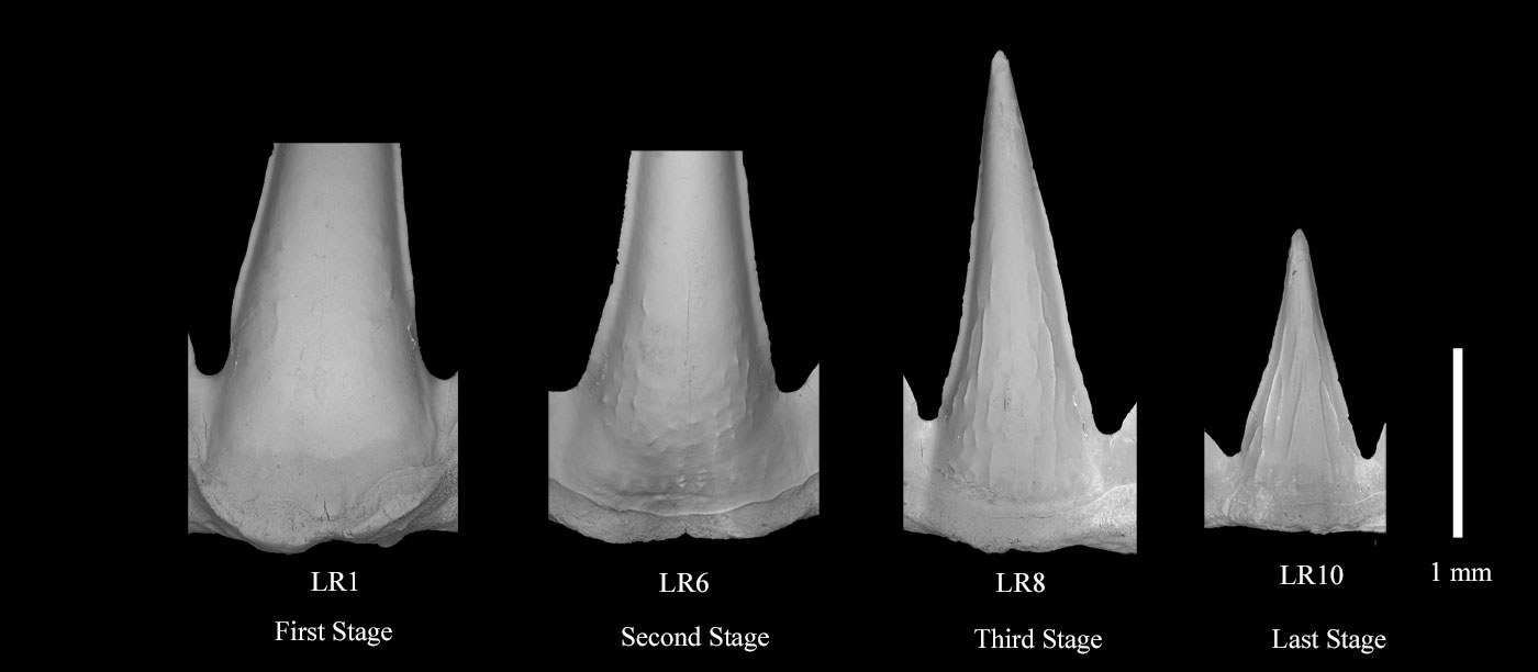
| Plate 1. shows the labial striae of the four teeth. |

| Differences in the morphology of the costule of each tooth from LR1 to
LR10 With the exception of the lowest ornamentations, the characteristics of each of the four tooth costule morphologies selected from the right side dentition of the lower jaw, 1. LR1; no stria observed. 2. LR6; short striae around 0.1 mm in length or shorter appear in the lower and middle portions of the tooth. 3. LR8; short striae are observed to be connected. Some short striae remain. 4. LR10; Almost all short striae have disappeared, forming long, connected striae. These have developed to near the top. |
| The costule Formation Process The differences in the stria morphology observed in the four teeth of the shark in this study can be considered as differences in the developmental stages of the stria, respectively. It can be inferred that this stria development process is similar in the teeth of other shark species. |
| Ref. 1. *1; Chlamydoselachus |
| Ref. 2. Four lower teeth of the frilled shark |

